Corneal Ulcers (Keratitis) in Knoxville: Our Ultimate Guide
The cornea is an important part of the eye that helps focus your vision as well as protects your eye from external dangers. Unfortunately, it can become damaged and develop corneal ulcers (keratitis) — open sores on the cornea. Being able to spot the signs can alert you to seek treatment for corneal ulcers in Knoxville.
In this blog we will cover:
- What is a corneal ulcer?
- Types of corneal ulcers
- Corneal ulcer symptoms
- Risk factors for keratitis
- Diagnosing corneal ulcers
- Corneal ulcer treatment in Knoxville, TN
- Corneal ulcer prevention
What Is a Corneal Ulcer?
Also known as keratitis , a corneal ulcer is an open sore on the cornea of the eye. They can develop for a number of different reasons, but they all have the same result: impaired vision. They do this by covering the cornea as well as the pupil of your eye much like crystal covers the face of a watch. This makes it much more difficult to see and can even lead to further issues.
Watch our AMA session with Dr. Janis Holt below to learn about corneal eye disease and other cornea problems!
What Does the Cornea Do?
The cornea acts as the “window” to the rest of your eye. Made of durable and transparent tissue, it works with your sclera (the white part of your eye) to help protect your eyes from:
- Dirt
- Germs
- Other harmful material
The cornea also acts as a filter, protecting your eyes from ultraviolet light.
Another important role it plays is in your ability to see. The cornea is slightly curved so that light is bent, or refracted , as it enters the eye. This allows you to focus on objects that are far away or close up.
This important part of the eye is made up of five layers . They are the:
- Epithelium
- Bowman’s layer
- Stroma
- Descemet’s membrane
- Endothelium
The epithelium comprises the outer layer of the cornea. It’s responsible for stopping any external particles from getting into the eye, whether it’s bacteria or dust. It also absorbs nutrients and oxygen from tears. The Bowman’s layer is a thin layer of connective tissue between the epithelium and the next layer, the stroma.
The stroma is the second thickest layer of the cornea following the epithelium. It’s composed of protein and water which gives it a solid but elastic density. The stroma is responsible for the domed shape of the cornea. Next is Descemet’s membrane . This thin layer separates the stroma from the final inner layer of the cornea known as the endothelium .
The endothelium is a single layer of cells that separates the stroma from the aqueous humor (the clear fluid in the front of the eye). The endothelium acts as a pump, removing any extra water that could not be absorbed by the stroma.
What Does the Pupil of the Eye Do?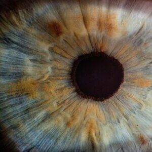
Corneal ulcers can also cover the pupil of the eye so it’s a good idea to understand what it is and the role it plays in your vision. Where the cornea is known as the “window” of the eye, the pupil is often referred to as the “aperture.” This term is taken from the part of a camera where light enters. The pupil serves the same function for your eye.
The pupil works with the iris to let in and control the amount of light that enters your eye. In fact, the size of the pupil is actually controlled by muscles within the iris, causing it to contract in bright conditions or dilate in low-light environments. Pupils also get smaller when focusing on close-up objects and grow larger when trying to see something that is far away.
Together, the cornea and pupil along with the iris absorb and control the light that enters your eye. As light passes through these and other parts of your eye, they eventually make it to your brain where they become the images that you see. Corneal ulcers impair this process from the very start, reducing your vision and making it difficult to see under any circumstance.
Click here to read more about corneal ulcers!
Types of Corneal Ulcers
Although they affect your eyes in similar ways, corneal ulcers actually come in a variety of types. This includes:
- Bacterial keratitis
- Fungal keratitis
- Acanthamoeba keratitis
- Herpes simplex keratitis
Bacterial Keratitis
Bacterial keratitis is caused by bacterial infections. It’s most common among those who wear contact lenses, but it can also occur in those who don’t wear them. Common types of bacteria that cause this form of keratitis include Pseudomonas aeruginosa and Staphylococcus aureus . Fortunately, bacterial keratitis can’t be spread from person to person.
Bacteria is everywhere and a common part of nature, your daily environment, and even your body. However, the bacteria that cause bacterial keratitis tend to be found in specific locations. Pseudomonas aeruginosa is commonly found in the water and soil you come into contact with. Staphylococcus aureus can be found on your skin and in the mucous membrane of your body.
Fungal Keratitis
Where bacterial keratitis is caused by a bacterial eye infection, fungal keratitis is caused by fungal infections. Common keratitis-causing fungi include:
- Fusarium species
- Aspergillus species
- Candida species
Fusarium and aspergillus are found in the environment. Fusarium is common in the soil, air, as well as on plants. Aspergillus is also found on plant material, including trees and grain crops. Candida , on the other hand, prefers to live on your skin and on your body’s mucous membrane.
Fungal keratitis is most common in tropical and subtropical climates, but it has been found in areas with milder temperatures. As with bacterial keratitis, it can not spread from person to person.
Acanthamoeba Keratitis
Acanthamoeba keratitis is a rarer form of the condition but poses significantly more danger to your eye health. It’s caused by free-living, microscopic single-celled organisms known as Acanthamoeba . This type of ameba is usually found in bodies of water such as oceans and lakes as well as in air and soil. This is why it is important not to swim with contact lenses in
Herpes Simplex Keratitis
Herpes simplex keratitis is caused by the herpes simplex virus. This type of keratitis usually heals without damage to the eye, but that doesn’t mean it can’t harm your eye health. Severe cases can lead to scarring of the cornea or even blindness. It can also predispose you to worse bacterial infections. It is a leading cause of blindness as well as the cause of most corneal infections around the world.
Unlike the previous types of corneal infections we’ve discussed so far, herpes simplex keratitis can be spread from person to person. It’s usually spread through the mouth, although it can spread through other parts of the body. Bouts of this form of keratitis usually occur when the earlier infection reactivates or “flares up.”
The herpes simplex virus, or HSV-1 for short, that causes herpes simplex keratitis is the same type of infection that causes cold sores on the mouth. While having HSV-1 does not necessarily mean you will develop this specific form of keratitis, some people are at higher risk than others.
This includes those who are:
- Female
- Non-Hispanic black or Mexican Americans
- Born outside of the United States
- Sexually active
Other Causes of Keratitis
Not all cases of corneal ulcers are caused by eye infections. They can also be caused by:
- Corneal abrasions
- Dry eye syndrome
- Bell’s palsy
Corneal abrasions are scrapes and scratches on the cornea. This can occur in a number of ways with anything coming in contact with your eye being able to cause corneal injuries. Having dry eye syndrome increases the chance of corneal abrasions. Dry eye syndrome is caused by your body not producing enough tears to lubricate your eyes to keep them comfortable and healthy.
Watch our AMA session with Dr. Erik Sweet below to learn about dry eye syndrome!
Bell’s palsy is a muscle disorder that causes weakness or paralysis in the facial muscles. Damage to the facial nerve results in symptoms starting suddenly and progressing over 48 hours. The paralysis caused by Bell’s palsy can prevent the eyelid from functioning properly and cause the cornea to dry out, leading to a corneal ulcer.
Corneal Ulcer Symptoms
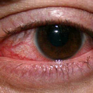
Symptoms of corneal ulcers can vary widely. Common symptoms include:
- Severe eye pain or soreness
- White spots in your cornea
- Sensitivity to light
- Discharge such as pus
- Feeling like something is stuck in your eye
- Redness of the eye
- Swollen eyelids
- Blurry vision
- Tearing
It’s important to keep in mind that many of these symptoms aren’t related specifically to the condition. Contact your Knoxville ophthalmologist immediately if you begin to experience any of these symptoms. Our eye doctors can diagnose the problem and provide you with the treatment you need to protect your vision.
Risk Factors of Keratitis
 Corneal ulcers can develop for a number of reasons with some people being at a higher risk than others. You may be more likely to develop one if you:
- Wear contact lenses
- Sleep in your contact lenses
- Have dry eye syndrome
- Use steroid eye drops
- Have an eyelid disorder that prevents it from functioning properly
- Have had a corneal abrasion or burn
- Have had cold sores, chickenpox, or shingles in the past
Having experienced one or more of these symptoms is not a guarantee that you will develop a corneal ulcer. However, they can be an indicator that you’re at a much higher risk. Talk to one of our ophthalmologists about your risk factors to learn if corneal ulcers are something you should be concerned about and what you can do about them.
Diagnosing Corneal Ulcers
Diagnosing corneal ulcers starts with an eye exam . It can be uncomfortable due to the open sore making it difficult to open the affected eye. However, testing the sharpness of your vision ( visual acuity ) is essential when making a diagnosis. This helps prevent and track the improvement of your corneal ulcer as it heals
Slit lamp exams are used when testing for corneal ulcers. A slit lamp works by providing a powerful light source and magnification, allowing your eye doctor to detect the extent and character of a corneal ulcer. This also allows them to see if it’s affecting any other structures within the eye.
During your exam, your eye doctor may use a penlight to test your pupil for reaction, size, and symmetry. They may also apply a special stain to your eye’s surface to determine the extent of the ulcer and to spot other irregularities. Tear or cell samples may also be taken and sent to a lab to learn the cause of the ulcer and to determine the best course of treatment.
To learn why comprehensive eye exams are vital for your eye health, click here !
Corneal Ulcer Treatment in Knoxville, TN
The form of treatment you receive will depend on the type of corneal ulcer that you have and its severity. Noninfectious and infectious forms of keratitis can be treated differently, and the treatments for infectious keratitis can vary depending on whether it’s bacterial, fungal, parasitic, or viral.
Noninfectious Keratitis Treatment
Your eye doctor will determine the severity of your corneal ulcer to decide which course of treatment is best for you. Artificial tear eye drops may be all that’s necessary in the case of a corneal scratch. More severe cases that cause significant pain and tears may require you to wear a 24-hour eye patch and use topical medications to heal your eye.
Bacterial Keratitis Treatment
Mild cases of bacterial keratitis due to Pseudomonas aeruginous or Staphylococcus aureus can often be treated with antibacterial eye drops. Moderate to severe infections may require oral antibiotics to fully remove the infection and restore your immune system. Your eye doctor will determine the severity of your infection during your exam and decide which is best for you.
Fungal Keratitis Treatment
Cases of keratitis due to a fungal infection are usually treated with antifungal eye drops and antifungal oral medications. This treatment can last for several months to ensure that the infection is fully removed from your body. Unfortunately, sometimes eye drops and oral medications aren’t enough. In these cases, corneal transplant surgery may become necessary.
Click here to read our article on corneal transplants and what to expect!
Parasitic Keratitis Treatment
The acanthamoeba parasite can be especially difficult to treat and remove from your system. Your doctor will prescribe antibiotic eye drops, in the beginning, to try and get rid of the parasite, but this doesn’t always work. As with fungal keratitis, severe cases may end up requiring surgery in order to cure you of your condition and restore your eye to normal.
Viral Keratitis Treatment
Viral cases of keratitis due to infections from herpes simplex keratitis can be treated in a number of different ways. Oral antiviral medications and antiviral eye drops are effective for many patients suffering from the condition. Infections from other viruses may be treatable through other means such as artificial tear drops.
Corneal Ulcer Prevention
If you wear contacts then taking them out and cleaning them regularly is of the utmost importance. Many cases of corneal ulcers are the result of poor contact lens hygiene, meaning that they can often be avoided with proper wear and cleaning. Keeping your lenses clean as well as your hands and the skin around your eyes is the best way to help prevent these ulcers.
When using soft and extended-wear contact lenses, make sure that you:
- Always wash your hands before touching your eyes or handling your contacts
- Use commercial contact lens cleaning solution, not homemade
- Clean and sterilize your lenses before and after you wear them
- Store them in disinfecting solution
- Don’t use water or saliva to store or moisten your lenses
- Wear your lenses as prescribed and not for prolonged periods of time
- Never wear your lenses overnight
To read our article on the dangers of wearing contact lenses overnight, click here !
Other tips for preventing keratitis include:
- Washing your face every night before bed, especially if you wear makeup
- Wearing protective eyewear during activities or work that could lead to an injury
- Keeping your eyes lubricated with artificial tears or ointment if you have dry eye syndrome
- Seeing your eye doctor immediately if you have symptoms of an eye infection
Water and Contact Lens Safety
Water can be a particular danger if you wear contact lenses. You may think your eyes are safe if you leave them in while swimming or showering, but water can carry bacteria that can be absorbed by your lenses and transfer them to your eyes. This is actually one of the main ways to contract acanthamoeba, so be aware of the dangers of exposing your contacts to water.
Click here to read more about water and contact lens safety!
To protect your eyes from bacteria and other harmful organisms in water, make sure that you:
- Take your contacts out and safely store them before swimming or showering
- Rinse your eyes once you’re done to get rid of any bacteria or chemicals
- Wear water-tight goggles if you must contacts while swimming and clean your lenses after you’re done
Following these tips will help you have a worry-free summer as you enjoy the water and keep your eyes healthy.
Are you concerned you may have a corneal ulcer in Knoxville, TN? Contact us today to schedule your appointment!
Corneal ulcers are sores that develop on the cornea. They are usually the result of different types but can have other causes. Symptoms can vary with white spots on your eye being common. Risk factors can include having dry eye syndrome, a previous corneal abrasion, or an eyelid disorder. Diagnosis involves a comprehensive eye exam and treatment can range from antibiotics to surgery. You can help prevent corneal ulcers with good hygiene and being careful about exposing your eyes to water.
Baptist Eye Surgeons is an ophthalmological practice in Knoxville, TN, and Morristown, TN. Give us a call at 865-579-3920 for more information or to schedule an appointment .