What Ophthalmologists Look for When Diagnosing Glaucoma
Glaucoma
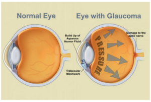 is an eye disorder that can occur when the pressure inside your eye rises above normal ( ocular hypertension) and damages your optic nerve. If left untreated, glaucoma can cause you to lose your peripheral vision and even go blind. Read more
about what causes glaucoma and how it affects your eyes.
is an eye disorder that can occur when the pressure inside your eye rises above normal ( ocular hypertension) and damages your optic nerve. If left untreated, glaucoma can cause you to lose your peripheral vision and even go blind. Read more
about what causes glaucoma and how it affects your eyes.
In fact, glaucoma is the second leading cause of blindness in the United States and affects more than 2.7 million people ages 40 and older. Because glaucoma presents with very few (if any) initial symptoms, nearly half of those who have glaucoma don’t even know it ( American Academy of Ophthalmology). This is one of the many important reasons to schedule an ophthalmologist appointment every year, especially if you’re over 40, are diabetic, and/or have a family history of glaucoma.
When conducting a comprehensive glaucoma exam, your doctor will test different parts of your eyes to look for any abnormalities in your inner eye pressure, the shape and color of your optic nerve, your complete field of vision, the angle in the eye where the iris meets the cornea, and the thickness of the cornea itself.
Gauging Your Inner Eye Pressure
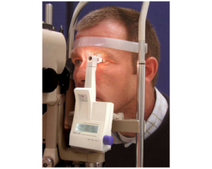 During tonometry, your ophthalmologist will measure the pressure within your eye with a tonometer. After applying numbing eye drops, he will then administer a small amount of pressure to your eye with a tiny device.
Normal eye pressure typically ranges from 12-21 mm Hg (a scale used to record eye pressure). Most glaucoma cases are diagnosed if the levels exceed 22 mm Hg, but because eye pressure is unique to each person, even those whose levels are as low as 12 mm Hg may be diagnosed with glaucoma as well.
Examining Your Optic Nerve
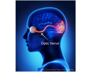 Your ophthalmologist will used a procedure called ophthalmoscopy to inspect your optic nerve for signs of damage caused by glaucoma. After dilating your pupils with eye drops, he will use special lenses to magnify your optic nerve and examine its shape and color.
A normal optic nerve is round and has a pink color around the rim. There is usually a central depression that is more yellow. An optic nerve that looks suspicious for glaucoma has a larger central depression (also known as the “cup”).
If your eye pressure is high and your optic nerve looks abnormal, your ophthalmologist may recommend one or more of the following diagnostic tests.
Mapping Your Complete Field of Vision
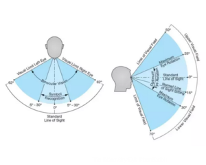 Perimetry is a visual field test that helps your ophthalmologist determine whether glaucoma has impacted your vision. During the exam, your ophthalmologist will ask you to look straight ahead and indicate when you see a light in your peripheral vision. Your ophthalmologist will then be able to draw a map of your complete field of vision using your responses. You may be asked to repeat the test to verify the results before your ophthalmologist makes a positive diagnosis.
If you are diagnosed with glaucoma, this test will be performed once or twice a year in addition to your regular eye exam to check for any changes in your vision.
Assessing Your Angle
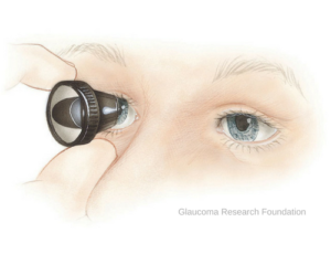 A gonioscopy is a fancy name for an exam that ophthalmologists use to measure the angle where the iris and the cornea meet. This angle will be described as open and wide or narrow and closed. After applying numbing eye drops, your ophthalmologist will gently place a special hand-held contact lens on your eye. The mirror on this contact lens will show your ophthalmologist whether the angle between the iris and the cornea is narrow and closed (signifying angle-closure, acute glaucoma) or open and wide (signifying open-angle, chronic glaucoma).
Measuring Your Thickness
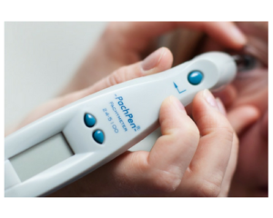 The thickness of your cornea can influence eye pressure readings, so your ophthalmologist may measure it using a pachymetry test. He can do this by simply placing a probe on the front part of your eye (the cornea) that will measure its thickness in under a minute—quick and painless. After knowing the thickness of your cornea, your ophthalmologist can better understand your intraocular pressure (IOP) readings and develop a treatment plan that’s right for you.
If you’re over 40, are diabetic, and/or have a family history of glaucoma and have not had a comprehensive eye exam this year, our ophthalmologists would be happy to see you. Just fill out this online form or call 865-579-3920 to schedule an appoint at our Knoxville or Morristown location.